
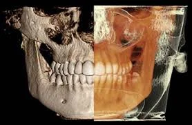
cone beam CT (CBCT) scan
Cone Beam Computer tomography (CBCT) London
This advanced technology generates a 3d scan of your tooth and related anatomy which provides us with valuable information to plan root canal therapy.
A routine x-ray is a 2d image which only shows the location of your teeth and the height of the bone. Cone Beam CT shows us cross-sectional views of your teeth in the jaw. The images it creates allow us to pinpoint difficult root anatomy, pathology relating to the tooth and much more. This helps simplify complex root canal procedures.
3d scanning is not needed in every case but becomes invaluable when more information is needed to complete cases successfully.
Advantages of Cone Beam CT (CBCT) for your treatment
The ability to demonstrates anatomy in 3 dimensions that intraoral and panoramic images cannot
Identify tricky root canal anomalies and determine root curvature
Diagnosis of dental periapical disease with no evidence of pathosis identified by conventional imaging
In cases where anatomic superimposition of roots or areas of the maxillofacial skeleton is impeding our diagnosis
Determine the extent of the periapical disease
In cases where complications have arisen to determine appropriate treatment planning to resolve such issues as:
1. overextended root canal obturation
2. material separated endodontic instruments
3. calcified canal identification
4. localization of perforations
Diagnosis and management of dentoalveolar trauma, root fractures, luxation, displacement of teeth, and alveolar fractures.
Diagnosis of internal/external root resorption or invasive cervical resorption
Surgical planning needed to determine the location of root apices and to evaluate the proximity of adjacent anatomical structures.